Stereotactic Radiosurgery
The Non-Invasive Neurosurgery

Stereotactic Radiosurgery
The Non-Invasive Neurosurgery
Introduction Evolution How Does it Work Indications Treatment Process Advantage
Frame Fixation Stereotactic Imaging Treatment Planning Radiation Delivery
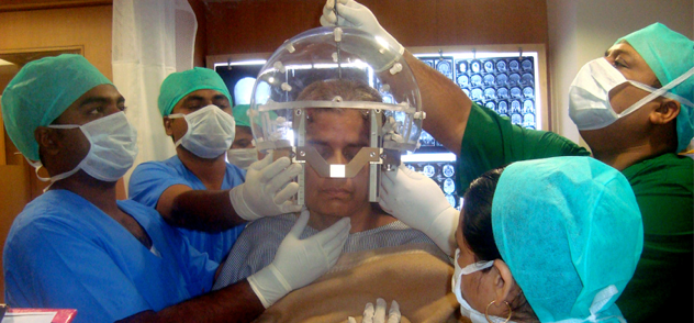
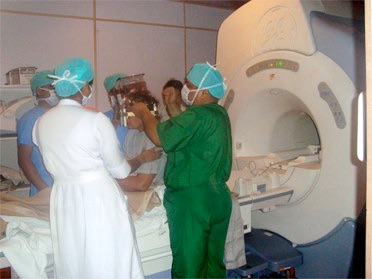
Stereotactic Imaging:
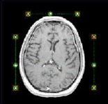
Images acquired with coordinate box show fiducial markers which help in registering image in the gamma knife workstation dedicated software. It is most important to get a distortion free image for good results as all treatment is to be done with guidance from these images.
Gamma Knife Perfexion as latest Gamma Plan PFX planning system which can define images more accurately then the previous versions. It also has capability to use images acquired before frame fixation and do a preplan before actual treatment day. Also it can used Images from PET Scan for better delineation for malignant tumors and assit in advanced planning. This software can also accept followup images to compare with treatment images and find change in size of tumor in follow-up.

Leksell Gamma Knife Perfexion
The Fifth Generation Robotic Gamma Knife